

How Digital Imaging Improves Dental Care is an important topic for anyone interested in modern dentistry and healthier smiles. Digital technology has changed how dentists examine teeth, identify hidden problems, and plan suitable treatments. Instead of relying only on traditional dental X-rays, professionals can use digital images, 3D scans, and intraoral cameras to understand a patient’s oral health more clearly. These tools make dental information easier to view, store, and share. They also help dentists explain their findings in a way patients can understand.
But digital imaging isn’t just about taking clearer pictures. It can support better treatment planning, more informed decisions, and improved communication during dental visits. From detecting cavities to planning dental implants, this technology offers several practical benefits. Understanding how it works can help you feel more confident about your next appointment.
What Is Digital Dental Imaging?
Digital dental imaging is the process of capturing and viewing images of teeth, gums, jawbones, and other oral structures using electronic equipment and computer software. Dentists use these images to investigate problems that may not be visible during a regular examination.
Unlike traditional film-based X-rays, digital radiographs can be displayed on a computer screen shortly after they are captured. Depending on the equipment, dentists may enlarge an image, adjust its display, or compare it with earlier records.
Digital dental imaging includes several technologies, such as:
- Digital dental X-rays.
- Panoramic dental imaging.
- Cone-beam computed tomography (CBCT).
- Intraoral cameras.
- Digital scans that create 3D models of teeth.
These tools don’t all work in the same way. Some use X-rays, while others capture visible-light images without ionizing radiation. A dentist chooses the appropriate method according to the clinical question and the patient’s needs.
Difference Between Digital and Traditional Dental X-Rays
Traditional dental X-rays use film that must be developed before the dentist can view the result. Digital X-rays use electronic sensors to capture images and display them on a screen.
| Feature | Traditional X-rays | Digital X-rays |
|---|---|---|
| Image capture | Dental film | Electronic sensor |
| Viewing | Film must be processed | Usually available quickly |
| Storage | Physical film records | Electronic records |
| Image adjustment | Limited | Some software adjustments available |
| Radiation exposure | Depends on equipment and technique | Often lower with comparable systems, but not always |
| Sharing images | Physical transfer or copies | Electronic transfer when secure and appropriate |
Digital technology offers convenience, but it doesn’t automatically guarantee a better diagnosis. Image quality, correct positioning, suitable exposure, and professional interpretation still matter.
How Digital Imaging Works During a Dental Visit
A typical digital imaging appointment follows a straightforward process:
- Step 1: Dental examination. The dentist reviews your symptoms, oral health history, and examination findings.
- Step 2: Image selection. If imaging is needed, the dentist chooses a suitable method.
- Step 3: Image capture. A sensor, scanner, or imaging machine captures the required information.
- Step 4: Image review. The dentist examines the results and looks for relevant signs of disease or structural changes.
- Step 5: Treatment discussion. The findings help guide recommendations, follow-up appointments, or additional tests.
Not every visit requires an X-ray or scan. The purpose is to obtain useful information when it can help with diagnosis or treatment.
What Are the Main Types of Digital Dental Imaging?
Different imaging technologies answer different questions. Understanding their uses can help patients see why a dentist might recommend one method rather than another.
Digital Intraoral X-Rays
Digital intraoral X-rays capture detailed images of individual teeth and the surrounding structures. A small sensor is placed inside the mouth while the X-ray machine captures the image.
Dentists may use these images to assess:
- Cavities between teeth.
- Tooth roots and surrounding bone.
- Changes around existing fillings or crowns.
- Certain infections or other abnormalities.
- Bone changes associated with gum disease.
Bitewing and periapical X-rays are common examples. Each provides a different view, depending on the area being examined.
Panoramic Dental Imaging
A panoramic X-ray captures a broad view of the teeth, upper and lower jaws, and nearby structures in a single image.
It may help dentists assess impacted teeth, developing teeth, jaw abnormalities, and certain problems that require a wider view of the mouth.
However, panoramic images generally don’t provide the same fine detail as some smaller intraoral X-rays. The dentist selects the method that best answers the clinical question.
Cone-Beam CT (3D Dental Imaging)
Cone-beam computed tomography, commonly called CBCT, uses X-rays and computer processing to create three-dimensional images of the teeth, jaws, and surrounding structures.
It may be useful for complex implant planning, impacted teeth, selected root canal problems, and certain oral surgical assessments.
CBCT isn’t necessary for every patient. It generally exposes patients to more radiation than conventional dental X-rays, so dentists should use it when the additional three-dimensional information is clinically justified.
Intraoral Scanners and Digital Dental Models
Intraoral scanners use optical technology to capture detailed surface images of the teeth and gums. Software combines these images into a digital model of the mouth.
Unlike X-rays and CBCT, an optical intraoral scan doesn’t use ionizing radiation.
These scans can support:
- Clear-aligner treatment planning.
- Digital impressions for crowns and bridges.
- Assessment of tooth shape and alignment.
- Communication with dental laboratories.
- Certain cosmetic dentistry procedures.
An optical scan shows surface details, not hidden structures inside the teeth or jawbone. It therefore complements other examinations rather than replacing every type of dental imaging.
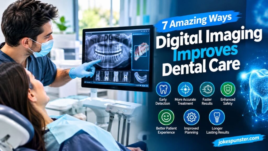
7 Amazing Ways Digital Imaging Improves Dental Care
Digital imaging can support several important parts of dentistry. Here are seven practical benefits, along with what patients should realistically expect.
1. Detecting Cavities and Dental Problems Earlier
Some dental problems develop without obvious pain or visible changes. A small cavity between two teeth, for example, may be difficult to identify during a visual examination alone.
Appropriate dental X-rays can help dentists investigate areas that are hard to see directly. Depending on the image and clinical findings, they may identify tooth decay, changes around roots, bone loss, or other abnormalities.
Earlier identification can create an opportunity to discuss treatment before a problem progresses. A small cavity may require less extensive treatment than decay that reaches deeper into a tooth.
Still, imaging doesn’t detect every dental condition. The dentist must interpret the image alongside symptoms, examination findings, and medical history.
2. Improving Treatment Planning and Clinical Decisions
Before beginning a dental procedure, a dentist needs a clear understanding of the affected area. Digital images can provide information that supports planning.
For example, a dentist considering an implant may need to assess bone dimensions and nearby anatomical structures. A CBCT scan may be appropriate when a two-dimensional image doesn’t provide enough information.
Digital images can also support decisions involving root canal treatment, impacted teeth, and complex restorative procedures.
The benefits include:
- Better visualization of relevant anatomy.
- More informed comparisons between treatment options.
- Improved preparation for selected complex procedures.
- Easier discussion of possible risks and limitations.
The right imaging method depends on the procedure. More detailed imaging isn’t automatically better if it doesn’t change the diagnosis or treatment plan.
3. Helping Patients Understand Their Oral Health
Dental explanations can sometimes feel confusing, especially when a dentist uses unfamiliar terms. Seeing an image of your own teeth can make the discussion easier to follow.
A dentist might enlarge an image to show a cavity, point out a change near a tooth root, or compare current findings with an earlier scan.
This visual approach can help patients:
- Understand why a treatment is recommended.
- Ask more specific questions.
- Compare suitable treatment options.
- Participate in informed decisions.
Digital images don’t replace a conversation with the dentist. They support it. Patients should still ask about the meaning of an image, the available options, and the likely benefits and risks of treatment.
4. Making Dental Records Easier to Access and Compare
Electronic dental records make it easier for practices to organize and retrieve images. When appropriate records are available, a dentist can compare new images with older ones to assess changes over time.
For instance, comparisons may help monitor certain bone changes, tooth development, or the condition of existing dental work.
Digital records can also support communication between general dentists and specialists. With suitable privacy protections, relevant images can be transferred securely when a referral is needed.
Useful records should be correctly identified, stored securely, and made available according to applicable privacy rules. Digital storage is convenient, but patient confidentiality remains essential.
5. Supporting More Precise and Conservative Treatments
Preserving healthy tooth structure is an important goal in modern dentistry. Digital images may help dentists understand the location and extent of a problem before deciding how to treat it.
For example, imaging can help assess decay or changes around a tooth root. During selected procedures, it can also provide useful information about the anatomy being treated.
This information may support a more targeted approach when clinically appropriate.
However, a digital image alone cannot guarantee a minimally invasive procedure, a pain-free experience, or a particular result. The outcome depends on the condition being treated, the dentist’s clinical judgment, and other patient-specific factors.
6. Improving Cosmetic Dentistry and Smile Planning
Digital photography, intraoral scans, and planning software can help dentists assess tooth shape, spacing, alignment, and smile proportions.
These tools may be useful when discussing veneers, crowns, clear aligners, or other cosmetic procedures. In some cases, software can provide a preview of a proposed change to help the patient and dentist discuss expectations.
Digital previews are planning aids, not guarantees of the final result. The actual outcome depends on oral health, the chosen procedure, materials, and clinical limitations.
Patients should discuss the appearance they want, the amount of tooth preparation involved, maintenance requirements, and possible alternatives before choosing cosmetic treatment.
7. Supporting Preventive and Personalized Dental Care
Every patient’s oral health needs are different. Age, dental history, symptoms, existing restorations, and risk factors can all influence the type of examination required.
Digital images can provide useful information when a dentist needs to monitor a particular concern or investigate a possible problem.
For example, suitable images may help assess changes between teeth or around supporting bone. Comparing records over time may also help the dentist decide whether further evaluation is necessary.
Prevention still depends on daily habits and regular professional care. Brushing with fluoride toothpaste, cleaning between teeth, maintaining a balanced diet, and attending recommended dental appointments remain important.
Digital imaging supports preventive care when it answers a relevant clinical question; it isn’t a substitute for these habits.
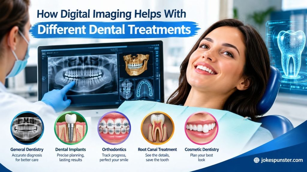
How Digital Imaging Helps With Different Dental Treatments
The usefulness of digital imaging depends on the treatment being considered. Some procedures benefit from detailed 3D information, while others rely on digital surface scans or conventional dental X-rays.
Dental Implants and Oral Surgery
Dental implant planning may involve assessing bone dimensions, the position of nearby structures, and the condition of the treatment area.
A CBCT scan may provide useful three-dimensional information when the dentist determines it is necessary. Digital planning can also support selected guided surgical workflows.
Not every implant case requires CBCT. The dentist should consider whether the additional information is needed and whether another method could answer the clinical question.
Braces and Clear Aligners
Digital scans can create models of the teeth and bite. Orthodontists may use these models to assess alignment, plan tooth movement, and design customized clear aligners.
Dental X-rays may also be needed in selected cases to evaluate tooth roots, developing teeth, or other structures.
The appropriate combination depends on the patient’s age, dental development, treatment plan, and clinical findings.
Root Canal Treatment
Root canal treatment involves examining and treating the inner part of a tooth. Dental X-rays can help assess root anatomy and changes around the root tip.
In selected complex cases, CBCT may provide additional information when ordinary images are insufficient. Because CBCT involves additional radiation, its use should be justified by the clinical situation.
Imaging supports diagnosis and planning, but the dentist must also evaluate symptoms and clinical examination findings.
Crowns, Bridges, and Cosmetic Dentistry
Digital intraoral scans can capture the tooth surfaces needed to plan many crowns and bridges. The resulting models may be used in computer-aided design and manufacturing workflows.
This can reduce the need for traditional impression materials in suitable cases and make communication with a dental laboratory easier.
Dental X-rays may be used separately when the dentist needs to assess the tooth roots, supporting structures, or other hidden areas. Surface scans and X-rays provide different information, so one doesn’t always replace the other.
Is Digital Dental Imaging Safe?
Digital dental imaging is widely used, but safety depends on selecting the right examination and using appropriate techniques. Some technologies, including digital X-rays and CBCT, use ionizing radiation. Optical intraoral scanners do not.
The American Dental Association recommends that dental radiographs be selected according to each patient’s needs rather than taken automatically at every visit. The benefits and risks should be considered before imaging is performed.
Understanding Radiation Exposure From Dental X-Rays
Digital dental X-rays often use less radiation than comparable conventional film systems, although exposure varies with the equipment and technique. CBCT generally involves more radiation than conventional dental X-rays.
The important point is that different scans have different purposes and exposure levels.
Patients can ask their dentist:
- Why is this image needed?
- Will it change the diagnosis or treatment plan?
- Is an existing image suitable for use?
- Is there an alternative that provides the necessary information with less or no radiation?
These are reasonable questions, especially when several imaging options are available.
Why Dentists Choose Imaging Based on Individual Needs
A patient with tooth pain may need a different examination from someone receiving an orthodontic assessment or considering an implant.
Dentists consider factors such as symptoms, examination findings, dental history, age, and the specific clinical question.
Children need particular care because they are more sensitive to radiation than adults. Imaging should be selected and adjusted for the individual patient.
The goal is to obtain enough information to support care without exposing the patient to unnecessary imaging.
How Dentists Minimize Unnecessary Radiation Exposure
Dental professionals can reduce unnecessary exposure by following established safety practices.
These include:
- Ordering images only when clinically justified.
- Reviewing suitable existing images to avoid unnecessary duplication.
- Choosing an imaging method appropriate to the clinical question.
- Limiting the scanned area when possible.
- Using appropriate exposure settings and equipment.
- Following relevant radiation-safety standards.
Patients should share their dental care history and mention recent imaging when relevant. This helps the dentist decide whether new images are necessary.
What Are the Limitations of Digital Dental Imaging?
Digital technology offers useful advantages, but it has limitations. Understanding them helps patients maintain realistic expectations.
Why Images Cannot Replace a Complete Dental Examination
A digital image shows only the information captured by a particular technique. It may not reveal every problem or explain every symptom.
A dentist may also need to examine the teeth and gums directly, review the patient’s history, assess bite function, and ask about pain or sensitivity.
Some conditions require additional tests or specialist evaluation. A normal image doesn’t necessarily mean that every part of the mouth is healthy.
Digital imaging works best as part of a complete diagnostic process, not as a stand-alone answer.
Equipment Costs, Image Quality, and Professional Interpretation
Digital imaging systems can be expensive to purchase, maintain, and update. These costs may affect the services available at different dental practices.
Image quality can also be affected by movement, positioning, equipment settings, or technical problems. Software adjustments may make certain details easier to view, but they cannot reliably recover information that wasn’t captured.
Finally, an image must be interpreted in the context of the patient’s condition. Technology can provide detailed information, but trained dental professionals remain responsible for diagnosis and treatment decisions.
The Future of Digital Imaging in Dentistry
Digital dentistry continues to develop as imaging, scanning, and treatment-planning systems become more connected. These improvements may make some dental workflows more efficient and help clinicians manage information more effectively.
Artificial Intelligence and Dental Image Analysis
Artificial intelligence software can assist with selected image-analysis tasks, such as highlighting areas that may need closer examination.
These systems may help dentists review information, but they can also make errors or miss important findings. Their output should not be treated as a confirmed diagnosis without professional evaluation.
AI is best understood as a support tool. A qualified dentist must consider the patient’s symptoms, history, examination results, and the limitations of the software.
Connected Digital Workflows and Personalized Treatment
Digital scans, imaging systems, design software, and dental laboratory tools can work together in some practices.
For example, a scan may support the design of a crown, while appropriate radiographs provide information about the underlying tooth and surrounding structures.
Connected workflows may reduce repeated steps and help teams share relevant information more efficiently. Secure handling of patient records remains important as more information is stored and transferred electronically.
The aim isn’t simply to use more technology. It’s to use suitable technology to improve patient care.
Frequently Asked Questions About Digital Dental Imaging
What is digital dental imaging?
it uses electronic imaging systems and digital scans to help dentists examine teeth, gums, jaws, and other oral structures.
Is digital dental imaging better than traditional X-rays?
It offers faster viewing and easier storage, and many digital X-ray systems use less radiation, but image quality and clinical suitability still matter.
Does digital dental imaging use radiation?
Digital X-rays and CBCT use ionizing radiation, while optical intraoral scanners capture surface images without it.
Can digital imaging detect cavities early?
Appropriate dental X-rays can reveal some cavities that aren’t easily visible during a regular examination, including decay between teeth.
Is 3D dental imaging safe?
CBCT can be appropriate when clinically necessary, but it generally uses more radiation than conventional dental X-rays and should not be used routinely without justification.
Conclusion
Digital imaging has improved modern dental care by giving dentists useful information for diagnosis, treatment planning, and monitoring oral health. Digital X-rays can help identify certain hidden problems, while 3D imaging and intraoral scans support specific procedures that require detailed views of teeth and surrounding structures.
The technology can also make dental records easier to manage and help patients understand their treatment options. However, it has limitations. Images must be interpreted by trained professionals, and radiation-based examinations should be used only when clinically justified.
Ultimately, the greatest value of digital imaging comes from combining appropriate technology with professional judgment, preventive care, and clear communication. If your dentist recommends an X-ray or scan, ask what it will help determine and how the results may affect your care. An informed conversation is a good step toward a healthier smile.